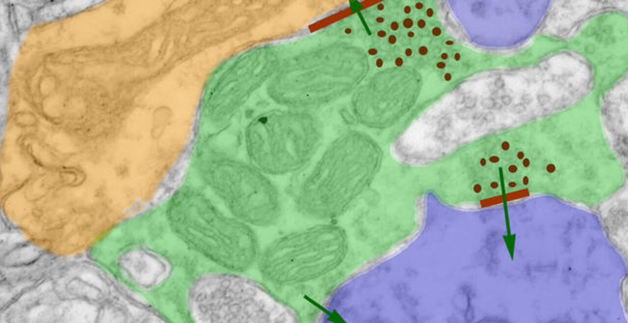
Synaptic connections among neurons that make up sensory pathways and other functional circuitries are the building blocks for how our brain functions, or in other words, how we see, hear, taste, move, learn, make plans or remember. These connections are amanable to change throughout life, allowing us to learn new skills, or to adapt to any external or internal alteration, including as we grow or age. By studying the morphological properties of neurons, synapses and identified axons, we aim to understand the synaptic inputs that converge on functionally distinct brain nuclei, how those inputs develop to form functional circuitries of the adult brain, and how they re-wire or degenerate.
What are the mechanisms by which the critical period of developmental plasticity is initiated, and terminated? Are there changes in the neurotransmitters, neuromodulators, hormones or their receptors that can signal for the changes in sensory perception or the behavior? How do glial cells interact with neurons in developing or aging brains? Using anatomical techniques including immuno-electron microscopy, ultrastructural morphometry, tract-tracing and confocal microscopy, we study synaptic circuitries in visual and gustatory sensory pathways during postnatal development, healthy adulthood and aging stages of our life span.
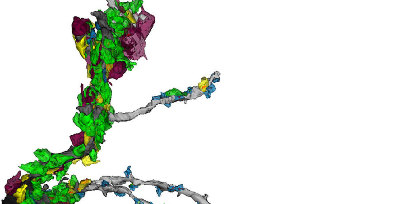
More to come: Morphology and Connectivity of Geniculate Relay Cell Dendrites and Synaptic Inputs in P14 Mice. Lead: Sydney Holton
Ultrastructural Neuropathology
Oligodendrocyte Dysregulation in Transgenic Alzheimer's Mice Models
Dysregulated oligodendrocyte and myelin dynamics as an early pathological feature of neuropil degeneration in Alzheimer's disease: an ultrastructural study. Erisir A, Maher EE, Anderson Z, Chawla S, Hanley L, Zhao A, Birisik K, Toklucu ES, Keskinoz EN.
I received an MD from Istanbul University School of Medicine in 1986 and a PhD in Behavioral Neuroscience from the State University of New York at Stony Brook in 1996. After my postdoctoral training at NYU Center for Neural Science and New York Medical School, Department of Physiology, I moved to the University of Virginia as an Assistant Professor in the Department of Psychology in 2000. I became a tenured Associate Professor in 2007 and Professor in 2013. I served as the director of undergraduate studies for the Cognitive Science (2008-2011) and the Neuroscience (2012-2015) programs, and as the Chairperson of my department (2016-2022).
My lab is equipped as a systems neuroanatomy, electron microscopy, quantitative morphology, and connectomics facility. The courses I teach at the undergraduate and the graduate level include Neural Mechanisms of Behavior (PSYC 4200); Psychobiology Lab (PSYC3210); Forum on Professional Conduct and Scientific Ethics (PSYC8040); Neural Mechanisms (PSYC7200); Neuropsychopharmacology (PSYC5285); Functional Neuroanatomy (PSYC5200); and Fundamentals of Neuroscience (PSYC 3200).
Contact Information
380A Gilmer Hall (office)
378/374 Gilmer Hall (lab)
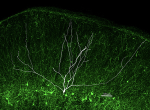
Wide-field (WF) neurons of the tectopulvinar pathway integrate retinal and cortical inputs via large dendritic arbors crucial for rapid visual motion detection. Previous studies identified potential marker genes for mouse WF neurons. Here, we validate CBLN2 as a molecular marker of the tree shrew WF neurons and construct AAVs that exploit CBLN2 promoter to selectively target WF neurons across species. Using intersectional genetics in the tree shrew, we show that WF neuron dendrites receive a distinct pattern of VGluT1+ and VGluT2+ inputs based on their distance from the cell body in the dorsoventral axis of the superior colliculus (SC). This represents the first example of a viral tool derived from the tree shrew genome for cell-type-specific targeting across species. Our results provide a foundation for studying SC circuitry in higher-order mammals and for extending this approach to additional conserved cell types in the SC and other brain regions.
Alzheimer's disease (AD) is a progressive neurodegenerative disorder traditionally defined by the accumulation of amyloid‑β plaques and neurofibrillary tangles. Increasing evidence suggests that white‑matter degeneration and myelin disruption occur early in disease progression and may contribute to neuropathological vulnerability. Here, we performed ultrastructural analyses in the 3xTg and 5xFAD mouse models of AD across developmental stages (3-12 months of age), including ages preceding overt amyloid plaque formation or neuronal loss. We identify a spectrum of oligodendrocyte‑ and myelin‑associated abnormalities, including single‑membrane herniations, myelin outfolds, and ectopic myelination of neuronal processes, which are evident as early as 3 months of age and are frequently associated with altered neuropil architecture and incipient dystrophic neurite morphology. These malformations were confirmed to be oligodendrocyte‑derived through O4 immunolabeling. Collectively, our findings reveal early, widespread myelin‑associated ultrastructural alterations that form a consistent structural component of neuritic pathology in AD models. We propose that dysregulated oligodendrocyte membrane remodeling represents an early pathological feature of AD, providing a framework for future studies examining how glial pathology intersects with neuronal degeneration and plaque‑associated neuritic remodeling.
The aggregation of the microtubule-associated protein tau into oligomeric complexes is strongly correlated with the onset and progression of neurodegeneration in Alzheimer's disease (AD). Increasing evidence implicates nuclear membrane disruption in AD and related tauopathies; however, whether this is a cause or consequence of neurodegeneration remains unresolved. Here, we show that nuclear lamina disruption emerges at the early Braak stages, coinciding with the initial formation of pathological tau aggregates in post-mortem AD brain tissue. Using the tauopathy mouse model (P301S PS19), we demonstrate that oligomeric tau (oTau) directly binds to the Lamin B Receptor (LBR), inducing nuclear envelope invaginations as revealed by electron microscopy. These structural alterations are accompanied by chromatin remodeling and gene expression dysregulation. To dissect the underlying mechanism, we employed a light-inducible OptoTau system (4R1N Tau::mCherry::Cry2Olig) in human iPSC-derived neurons, enabling real-time visualization of tau aggregation dynamics. This system revealed selective recruitment of oTau to the nuclear envelope and direct interactions with LBR and Lamin B2, leading to nuclear deformation and activation of the protein translational stress response. Together, these findings identify nuclear membrane disruption as an early and potentially causative event in tau-mediated neurodegeneration, establishing a mechanistic link between tau oligomerization, nuclear stress, and chromatin remodeling. Targeting nuclear destabilization may offer new therapeutic avenues for mitigating AD pathogenesis.
The superior colliculus (SC), a midbrain sensorimotor hub, is anatomically and functionally similar across vertebrates, but how its cell types have evolved is unclear. Using single-nucleus transcriptomics, we compared the SC's molecular and cellular organization in mice, tree shrews, and humans. Despite over 96 million years of evolutionary divergence, we identified 30 consensus neuronal subtypes, including Cbln2+ neurons that form the SC-pulvinar circuit in mice and tree shrews. Synapse-related genes were among the most conserved in the SC, unlike neocortex, suggesting co-conservation of synaptic genes and collicular circuitry. In contrast, cilia-related genes diverged significantly across species, highlighting the potential importance of the neuronal primary cilium in SC evolution. Additionally, we identified an inhibitory SC neuron in tree shrews and humans but not mice. Our findings reveal that the SC has evolved by conserving neuron subtypes, synaptic genes, and circuitry, while diversifying ciliary gene expression and an inhibitory neuron subtype.


