Progression of Cellular Degeneration in Triple Transgenic AD Mice
In brains that will be diagnosed as with Alzheimer's Disease, the cholinergic cells of the basal forebrain are found to die first.
The same region displays the earlier signs of pending cellular death in 3xTg mice as young as postnatal month 3 (PM3).
PM3- Basal Forebrain

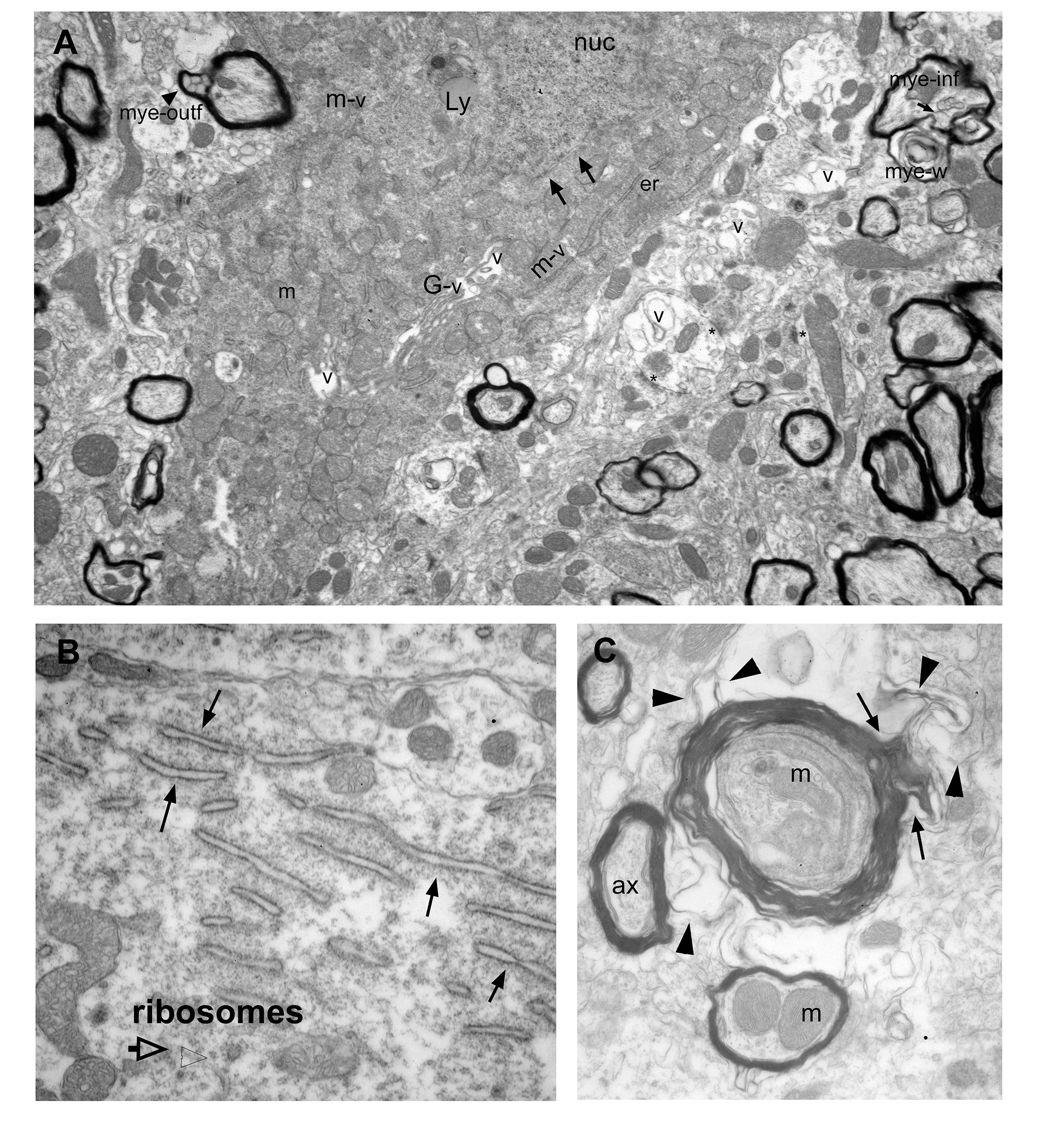
A. A cell body shows signs of cytoplasmic degeneration: Cytoplasm is electron dense; nuclear membrane is dissolved (arrows); endoplasmic reticulum (er) are sparse and display enlarged lumens; vacuoles are frequent and associated with cellular organelles including Golgi apparatus (G), mitochondria (m), and ER (er). Also note various myelination pathologies the surrounding neuropil, including myelin outfolds (mye-outf), myelin infolds (mye-inf), and myelin whorls (w). Abbrv: nuc: nucleus; Ly: Lysosome.
B. ER stress is characterized by the enlargement of its lumen (arrows). This cell is otherwise healthy, with free and ER-associated ribosomes int he cytoplasm.
C. Examples of myelination pathologies frequently encountered in the basal forebrain in PM3 brains. In this example, thick myleing surrounds a collection of neuropil components including membranes, mitochondria, and multivesicular bodies. The myelin sheet gives off a filamentous outfold (arrows), which invades the surrounding neuropil (arrowheads).
PM 5- Basal Forebrain

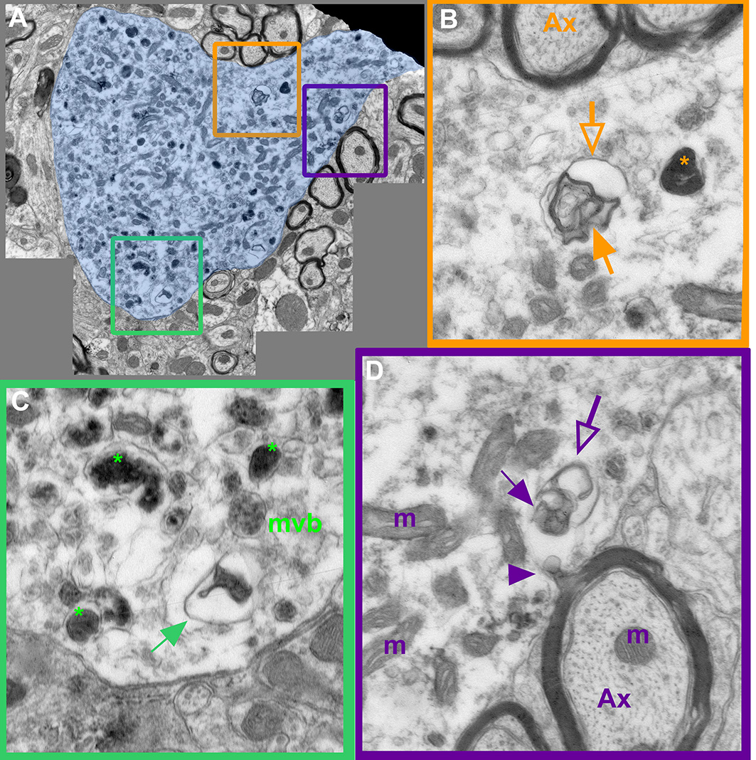
A. A large cell body cross-section, including a dendrite emergence point. Cytoplasm of this cell is not electron dense however, many degeneration products fill the cell, scattered among discernable mitochondria (m).
B, C and D are enlarged views of the regions in A, marked with orange, green and purple frames.

