Identified neurons in the medicinal leech Hirudo verbana
Dorsal Aspect
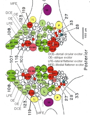
Identified neurons on the dorsal aspect of segmental ganglia in the leech, Hirudo medicinalis.
| Map numberc | Major axon projectionb | Color codea | Descriptionb | Behavioral Involvementbc |
| Motoneurons | ||||
| DE-3 | CPNR | green | excitor of DLM | swimming, LB crawling |
| DE-5 | CPNR | green | swimming, LB crawling |
|
| DE-18 | CANR | green | swimming | |
| DE-107 | CANR | green | swimming, LB | |
| VE-4 | IPNR | green | excitor of VLM | swimming, LB crawling |
| VE-8 | CPNR | green | swimming, LB | |
| VE-108 | CANR | green | swimming, LB | |
| FE-109 | CNR CNR |
green | excitor of lateral dorsal-ventral FM |
swimming, LB |
| FE-117 | CNR | blue | excitor of medial dorsoventral FM |
|
| DI-1 | CNR | red | inhibitor of DLM | swimming, LB crawling |
| DI-102 | CNR | red | swimming, LB | |
| VI-2 | CNR | red | inhibitor of VLM | swimming, crawling |
| VI-7 | CNR | red | swimming | |
| VI-119 | CNR | red | swimming | |
| FI-101 | CNR | red | inhibitor of dorsoventral FM |
swimming, LB |
| CE-11 | CNR | blue | excitor of CM | |
| CE-12 | CNR | blue | ||
| CE-112 | CNR | blue | ||
| L (Large LM neuron) | LM | blue | excitor of other | crawling |
| DE-6 | CNR | blue | local bending | |
| LI-9 | CNR | blue | inhibitor of LM | |
| LE-106 | CNR | blue | excitor of lateral LM | |
| OE-110 | CNR | blue | excitor or OM | local bending |
| OE-111 | CNR | blue | ||
| Interneurons | ||||
| 21 | green | serotonergic, gating | swimming | |
| 27 | AIC, one ganglion |
red | oscillator 240° phase |
swimming |
| 28 | AIC, one ganglion |
red | oscillator 120° phase |
swimming |
| 33 | AIC, one ganglion |
red | oscillator 240° phase |
swimming |
| 115 | PCC | red | oscillator 0° phase |
swimming, LB |
| 123 | PIC, one ganglion |
red | oscillator 0° phase |
swimming |
| 125 | PIC | blue | excitor, receives input from P cells |
LB |
| 34 | blue | interacts with T cells, S cells |
||
| Mechanosensory cells | ||||
| P | INR, IC | yellow | mechanosensory | responds to pressure |
| N | INR, IC | yellow | mechanosensory | responds to noxious |
| Neurosecretory cells | ||||
| 50 | IC | pink | secretary neuron | LB, feeding |
aColor codes correspond to the maps of segmental ganglia (Fig. 2). Coded with green and red are involved in swimming circuits.
bAbbreviations:
LM-longitudinal muscles
DLM-dorsal LM
VLM-ventral LM
FM-flattener muscles
CM-circular muscles
OM-oblique muscles
NR-nerve root
CANR-contralateral anterior NR
CNR-contralateral NR
CPNR-contralateral posterior NR
INR-ipsilateral NR
IPNR- ipsilateral posterior NR
IC-ipsilateral connectives
AIC-anterior IC
PIC-posterior IC
PCC-posterior contralateral connectives
LB-local bending
CM-circular muscle
NR-nerve root
CNR-contralateral NR
INR-ipsilateral NR
CANR-contralateral anterior NR
IC-ipsilateral connectives
AIC-anterior IC
PIC-posterior IC
CC-contralateral connectives
PCC-posterior CC
ACC-anterior CC
Fn-Favre’s nerve
cThis table provides an update of neurons listed in Muller KJ Nicholls JG Stent GS (1981) Neurobiology of the leech. Cold Spring Harbor, NY: Cold Spring Harbor Laboratory.
Ventral Aspect

Identified neurons on the ventral aspect of segmental ganglia in the leech, Hirudo medicinalis.
| Map numberc | Major axon projectionb | Color codea | Descriptionb | Behavioral Involvementbc |
| Motoneurons |
| Map numberc | Major axon projectionb | Color codea | Descriptionb | Behavioral Involvementbc |
|---|---|---|---|---|
| Motoneurons | ||||
| AE | CNR | blue | annuli erector | change skin surface from smooth to ridged input from T cell |
| CV | CNR | blue | excitor of ventrolateral CM | crawling |
| HE | INR (M3-19) | blue | cardiac rhythmic movement | control HN |
| AP | CNR | blue | input from P cell | position detection, crawling |
| 152 | CNR | blue | CM excitor | crawling |
| 166 | CNR | blue | CM inhibitor | crawling |
| Interneurons | ||||
| 61 | IC (2 ganglia) | green | serotonergic gating |
swimming |
| 204 | Fn (unpaired) | green | gating | swimming, crawling, behavioral decision |
| 205 | Fn (unpaired) found in M9 |
green | gating | swimming |
| 208 | IC (unpaired) | green | oscillator | swimming |
| 60 | IC (2 ganglia) | red | oscillation 240° | swimming |
| 151 | IC (NR) | blue | non-spiking | a negative feedback teo limit excessive motor activity, crawling |
| 153 | CANR | blue | receive excitatory photosensory input oscillation 180° |
photoreception, swimming |
| 154 | blue | photosensory input | photoreception | |
| 157 | CC | blue | EPSP long latency | LB |
| 159 | PIC | blue | EPSP long latency | LB, crawling |
| 160 | blue | shifts swim phase but no oscillation | swimming? | |
| 161 | IC | blue | EPSP from P cell | LB |
| 162 | IC | blue | EPSP from P cell | LB |
| 169 | CC | blue | EPSP long latency | swimming, LB |
| 201 | CC | blue | receive inhibition from the SMRs | water motion detection |
| 202 | CC | blue | receive excitation from the SMRs | water motion detection |
| 213 | AIC | blue | ascending | crawling |
| 215 | blue | receive excitation (ipsi-) sensillum 7 |
photosensory reception | |
| 216 | blue | receive photosensory inputs | photosensory reception | |
| 212 | ACC | blue | EPSP from P cell | LB |
| 218 | IC | blue | spotaneous EPSP | LB |
| 258 | INR | blue | with large soma | crawling |
| S | Fn | blue | LB, shortening, learning, crawling | |
| HN | IC (M1-M7) | blue | inhibitor IN heart oscillator |
control of heartbeat |
| E21 | Fn | M21 | trigger/gating | swimming |
| Mechanosensory cell | ||||
| T | IC (NR) (T1, T2, T3) |
yellow | responds to tactile stimuli | touch |
| P | IC (NR) (P1, P2) |
yellow | responds to pressing stimuli on skin | pressure |
| N | IC (NR) (N1, N2) |
yellow | responds to noxious stimuli on skin, gut | nociception |
| Neurosecretory cell | ||||
| R (Retzius) | ICNR, IC | pink | contains serotonin | swimming, feeding, muscle relaxation |
| 50 (Leydig) | pink | LB, feeding |
aColor codes correspond to the maps of segmental ganglia (Fig. 2). Coded with green and red are involved in swimming circuits.
bAbbreviations:
LM-longitudinal muscles
DLM-dorsal LM
VLM-ventral LM
FM-flattener muscles
CM-circular muscles
OM-oblique muscles
NR-nerve root
CANR-contralateral anterior NR
CNR-contralateral NR
CPNR-contralateral posterior NR
INR-ipsilateral NR
IPNR- ipsilateral posterior NR
IC-ipsilateral connectives
AIC-anterior IC
PIC-posterior IC
PCC-posterior contralateral connectives
LB-local bending
CM-circular muscle
NR-nerve root
CNR-contralateral NR
INR-ipsilateral NR
CANR-contralateral anterior NR
IC-ipsilateral connectives
AIC-anterior IC
PIC-posterior IC
CC-contralateral connectives
PCC-posterior CC
ACC-anterior CC
Fn-Favre’s nerve
cThis table provides an update of neurons listed in Muller KJ Nicholls JG Stent GS (1981) Neurobiology of the leech. Cold Spring Harbor, NY: Cold Spring Harbor Laboratory, Appendix Table 1
References for additions to appendix Table 1
Leydig cell Wilson RJ, Kristan WB Jr, Kleinhaus AL (1996) An increase in activity of serotonergic Retzius neurones may not be necessary for the consummatory phase of feeding in the leech Hirudo medicinalis. J Exp Biol 199:1405-1414
LB behavior Lockery SR, Kristan WB Jr. (1990) Distributed processing of sensory information in the leech. II. Identification of interneurons contributing to the local bending reflex. J Neurosci 10:1816-29
Garcia-Perez E, Zoccolan D, Pinato G, Torre V (2004) Dynamics and reproducibility of a moderately complex sensory-motor response in the medicinal leech. J Neurophysiol 92:1783-1795
Wittenberg G, Kristan WB Jr (1992) Analysis and modeling of the multisegmental coordination of shortening behavior in the medicinal leech. II. Role of identified interneurons. J Neurophysiol 68:1693-1707
AP cell Shan D, Zhang RJ (2001) Frequency coding of positional information by an identified neuron, the AP cell, in the leech, Whitmania pigra. Brain Res Bull 56:511-515
AE cell Rodriguez MJ, Iscla IR, Szczupak L (2004) Modulation of mechanosensory responses by motoneurons that regulate skin surface topology in the leech. J Neurophysiol 91:2366-2375
Cell 151 Wadepuhl M (1987) A morpho- and physiologically uncommon neuron in the leech CNS. Naturwissenschaften 74:43-45